|
Hemangioma
 Overview
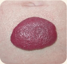
Hemangioma is the most common type of birthmark, occurring in 10–12% of all children by age one. It is a benign tumor of the cells, called endothelial cells, that normally line the blood vessels. In hemangiomas, the endothelial cells multiply at an abnormally rapid rate. Hemangiomas are not usually hereditary, although 10% of infants have a family history of these vascular birthmarks.
Most hemangiomas do not require any treatment because they go away on their own. In some children, loose skin, discoloration, or tiny, dilated blood vessels may remain after the hemangioma has fully involuted. When this occurs, hemangioma laser therapy will improve the child's appearance. Babies as young as several weeks of age can be effectively treated with the pulsed dye laser. What to Expect
Prior to the treatment, a topical anesthetic may be applied to the lesion, the child is immobilized, and his or her eyes are covered to shield them from the laser light. Depending on the size of the hemangioma, a treatment to an infant's face may last only 5–10 minutes. The child usually will only experience mild discomfort similar to repeated pinching or multiple snaps of a rubber band.
Immediately after treatment, the lesion turns purple-gray, which may darken over the next 24 hours and last for 10–14 days. Multiple treatments, spaced approximately two months apart, are usually necessary to attain the degree of lightening desired. The risks of scarring or permanent changes in the skin's pigmentation are very small. Occasionally, a hemangioma will ulcerate, creating painful sores that are difficult to heal. Although the pulsed dye laser can aid this healing process, multiple treatments of about once every 3 weeks are necessary because these lesions will continue to grow. Effectiveness and Results
Most hemangiomas, when first diagnosed, are only superficial; these can be treated with laser therapy right away. Early treatment is key, as laser becomes less effective if you wait. The laser selects the red area and shrinks the vessels so that the result is a less noticeable lesion. Repeated treatments can almost completely remove the superficial component. However, since the laser can only penetrate 1–3mm, it cannot shrink any deep component.
Sometimes, early treatment will prevent further growth, although deeper portions may still persist and grow. The risk of scarring is small. Realistically, the complete removal of every trace should not be expected. | |||